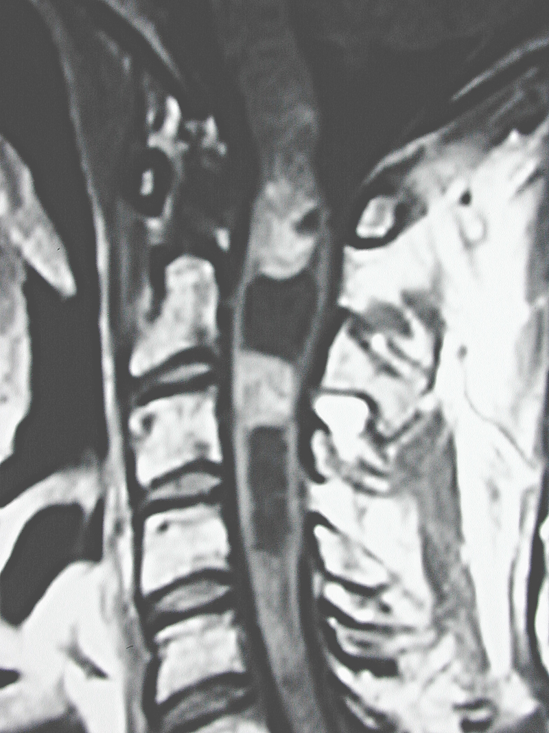
General Findings
- Spinal cord diameter: The diameter of the spinal cord will be enlarged. If it is not, a disease other than neoplasia should be suspected.
- Eccentric: The epicenter for tumor will be off the midline involving one side of the cord more than another to a variable degree.
- Smaller average length: The length of the spinal cord involved by the tumor is, on average 4 spinal segments.
- Malignant tumors: These tumors can appear similar to benign.
T1-Weighted Sequence
- Hypointense: 80% of time the tumor will appear slightly hypointense with the remaining 20% being isointense.
- Blurred margins: The margins of the tumor will appear slightly blurred due to the infiltrative nature of astrocytomas.
- Gadolinium enhances: In almost all cases there will be uptake of gadolinium but the degree will vary. The enhancement can be homogeneous or heterogeneous.

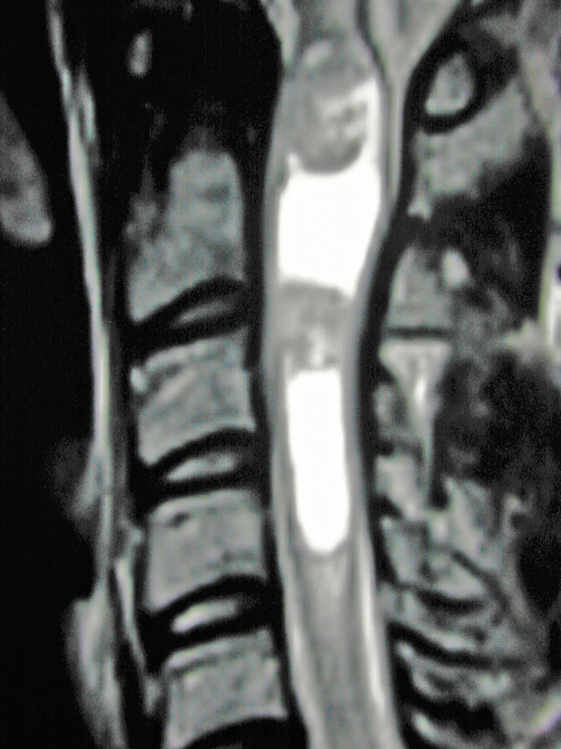
T2-Weighted Sequence
- Hyperintense: There is a variable degree of T2 hyperintensity in astrocytomas of the spinal cord.
- Nonhomogeneous: The degree T2 hyperintensity within the tumor will vary (as opposed to edema, which is homogeneous).
- Size matches T1-weighted sequence: The size of the tumor seen on T2 imaging will match that seen on T1 imaging with benign tumors.
Please create a free account or log in to read 'MRI of Intramedullary Spinal Cord Astrocytoma'
Registration is free, quick and easy. Register and complete your profile and get access to the following:
- Full unrestricted access to The ISPN Guide
- Download pages as PDFs for offline viewing
- Create and manage page bookmarks
- Access to new and improved on-page references